Metabolic Cancer Therapies
Put simply, cancer cells have aberrant energy metabolism compared to normal cells. Metabolic Cancer therapies attempt to exploit this difference. Cancer cells have a reduced capacity to generate energy with oxygen (oxidative energy production), with a concurrent increase in energy generation without oxygen. On Average a healthy cell produces 89% of its energy using oxygen, and 11% through non-oxidative metabolism (non-oxidative metabolism is also known as “fermentation”). Oxidative energy production is far more efficient than fermentation. Almost 20 times more energy is released when glucose is completely oxidized, as opposed to when it is fermented.
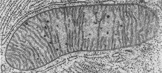
Healthy Mitochondria. Note the abundant looping structures inside the mitochondria (cristae), this is where all energy is produced through oxidative pathways.
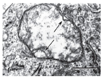
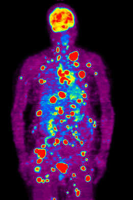
Oxidative energy production takes place in a cellular organelle called the mitochondria. The mitochondria are known as the cellular “power plants.” Also telling is the fact that the greater the degree of fermentation displayed by a given cancer, the more aggressive the cancer. Because a tumor cell’s mitochondria appear to be dis-regulated, and generate energy by such an inefficient pathway, they have to consume much more glucose to remain viable. A glance at a PET scan, which uses a radioactive labeled glucose analog to image cancer, provides stunning visual evidence of the voracious appetite tumor cells have for glucose compared to normal tissue.
Altered Metabolism May be One of the Drivers of Cancer
It is well established that once a cell has an impaired ability to produce energy through oxidative pathways, the genomic instability (increased potential for DNA mutations to occur) that accompanies tumor development, inevitable follows. While it’s true that most of the agents known to cause cancer; chemical carcinogens, viruses, radiation, and inflammation can cause mutations to DNA, it is also true these provocative agents damage the mitochondria. The metabolic theory of cancer states that once the mitochondria of a given cell become disregulated, and the cell reverts to fermentation to obtain energy, a plethora of metabolic and epigenetic modifications participate in the origin of cancer.


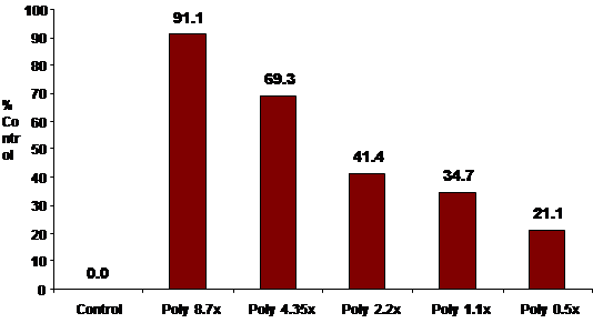
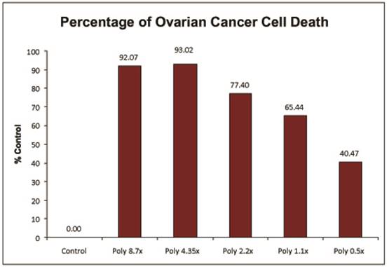
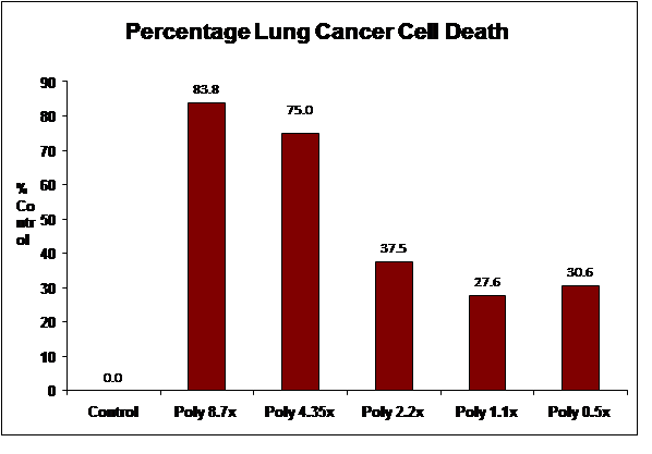
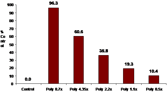
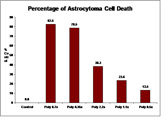
CANCER CELL DEATH RATE IN 5 CELL LINES
After 48 hours of exposure